3D high-tech diagnostics

20th anniversary of digital volume tomography (DVT) – Precise, safe, gentle & fast
“And now please open your mouth once a long way!” – What you then see as a dentist, is far from enough to plan with the naked eye a tailor-made dentures. How your jaw is made, shows in the first instance an x-ray. Normally, a two-dimensional panoramic image is taken, which allows the detection of caries or the localization of inflammation. However, it is not enough to make a detailed and meaningful diagnosis when z. B. implants are planned.
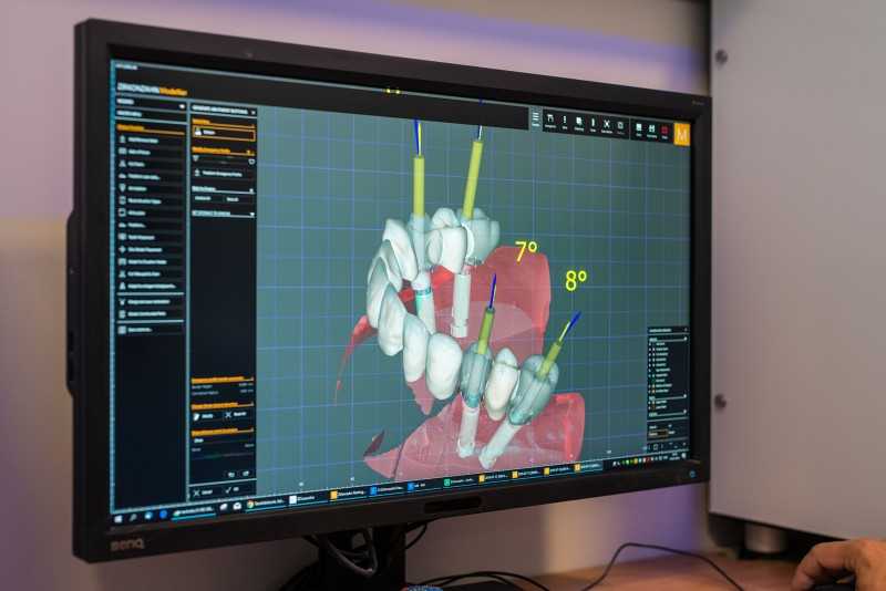
In such a case, the method of digital volume tomography (DVT) – usually called 3D X-ray – used. The DVT is a high-tech X-ray device that was specially developed for the head area and can be used to create three-dimensional images.
advantages
We can get a more accurate picture of the anatomical conditions, as the structures of the body are also displayed in depth. Furthermore, the recordings are particularly detailed, true to scale and undistorted. In general, treatments, such as implantation, can be planned more precisely and thus usually performed more safely and gently.
Often, for example, it is only possible through the 3D representation to recognize the exact width of the jawbone or to assess the spatial course of an important nerve tract and to protect it accordingly.
functionality
1. Creation of individual cross-sectional images of the jaw or of the entire head
2. Single images can be used directly for true-to-scale surveying or assembled using a computer program to a complete image of the jaw
3. Complete image is available, for example, for computer-assisted 3D implant planning
applications
Field of implantology
Planning of implantations as well as all jaw operations, which can be designed more successfully – distance to running nerves, maxillary sinus, adjacent teeth as well as the bone supply can be accurately measured
Area of periodontics
The extent and course of the bone loss can be determined exactly – this can be carried out targeted bone augmentation measures
Suspected herd in the tooth and jaw area
Root dead teeth, cysts, even the smallest root remains or amalgam tattoos in the jaw can cause harmful spill effects – even on the entire human organism
Production of high-quality dentures
Before the production must be partially clarified whether planned holding teeth are sufficiently strong to wear the dentures permanently and whether the investment in the dentures worthwhile
Recognition of temporomandibular joint disease
Comparison 2D X-ray, CT & DVT
In the conventional 2D X-ray method, the region to be imaged is transilluminated by an X-ray source and imaged on an X-ray film or sensor. On two-dimensional images, the structures can overlap and cover each other, whereas CBCT images provide three-dimensional images that allow us to see from different angles on the anatomical conditions.
Compared to the normal panoramic view, however, the radiation in the DVT can be increased up to ten times, depending on the device.
A computer tomograph (CT) provides – similar to the DVT – three-dimensional X-ray images. In contrast to a CT scan, where spirally thin axial layers are created, a cone-shaped X-ray rotates around the area to be detected during the DVT scan. Within a few seconds, a large number of two-dimensional individual images are generated. These individual images are combined by sophisticated reconstruction algorithms to a detailed three-dimensional data set (volume data set).
The DVT is in comparison to the frequently used computed tomography (CT) but significantly reduced in radiation.
 Source: DVT Academy Germany
conclusion
This 3D volume data set allows us to accurately analyze the – individually tailored – treatment region, providing detailed information on existing bone material, the course of nerves, the exact location of displaced teeth, the nature of paranasal sinuses, and the position of the hammer, anvil, stapes, and more.
This precise knowledge of anatomical conditions reduces the risk of misinterpretation and significantly increases the planning and treatment safety for you and us. Digital volume tomographs have become the basis for diagnostic and planning work in dental surgery.
With this high-tech diagnostic tool we can plan the interventions for you with unprecedented safety!
We are happy to help you with further questions about 3D X-rays. Just make an appointment!
Related Posts
-

High-tech in dentistry, gelencsér dental
High tech in dentistry In our clinic, the treatments are carried out with the most modern dental technology, the diagnostic work, the practical…
-

implants We predominantly use full titanium screw implants that have been in clinical use for decades (clinical use on patients began in 1965). For each…
-

Dental Implants: Costs, Manufacturers, Experiences and Treatment
Everything about dental implants There are certainly many questions that you would like clarified if you lose one or more teeth and this through Dental…
-

functional analysis The detail measurement (facebow) For the initiation of the correct treatment of a temporomandibular joint disease, as well as for the…
