miosis
The miosis is the bilateral constriction of the pupils in the event of incidence of light or during near-fixation. If a miosis is present without a light stimulus and independent of the near fixation, this phenomenon has disease value. Intoxications come as a cause in question, such as meningitis or lesions of the pons.
Table of Contents
What is a miosis?
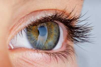
In the case of miosis, the pupils temporarily narrow to up to two millimeters. The constriction may be either one or both sides pronounced and have different strength. The reflex corresponds to an eye reflex to light irradiation and is subject to parasympathetic control.
Either the constriction results from a contraction of the vegetatively controlled ocular muscle sphincter pupillae muscle or by a reduced activity of its antagonist dilator pupillae muscle. Both muscles are part of the inner eye muscles.
Miosis can be the symptom of various diseases. But it can also be triggered artificially by the administration of parasympathomimetics. The opposite of miosis is the mydriasis, in which there is a dilation of the pupils over five millimeters.
The lens constriction and lens extension both belong to the phenomenon of accommodation. They are physiological in response to certain stimuli. Without previous stimulus, however, it is pathological phenomena.
function & task
The parasympathetic fibers of the third cranial nerve are interconnected in the ciliary ganglion, a ganglion in the eye socket responsible for pupillary reflexes. The nerve fibers extend through the Nervi ciliares breves to the Musculus sphincter pupillae.
The reflex arc of the pupillary reflexes starts at the retina (retina). Via the optic nerve it continues into the area pretectalis and is connected on both sides in the mesencephalon. In a physiological miosis, as is the case with light stimuli, this bilateral interconnection always narrows the pupils on both sides. This is true even if only one eye is directly irritated. The other eye is then talked about an indirect light reflex.
The adaptation to the incidence of light is referred to as adaptation. The narrowness reduces the incidence of light and the eye thus preserves the visual acuity. Miosis is thus both a protective reflex and an adaptation reflex.
Physiologically, in the broadest sense, near-fixation also leads to miosis. Together with the convergence movement and the accommodation, the miosis in the near-fixation makes up the neurophysiological control loop of the near-focus stria. The narrowing of the pupil as part of the accommodation helps people to see nearby objects particularly sharply, because the reduced lens generates a higher depth of field. Even in lensless people, miosis improves visual acuity. Therefore, it is targeted and deliberately brought about by the ophthalmologist for the treatment of various diseases in order to improve the eyesight of the patients.
Here you will find your medication
Diseases & complaints
The miosis can be brought about deliberately by medication and then usually corresponds to a ophthalmological therapy, as it may be useful, for example, in glaucoma. Targeted induction usually takes place with miotics such as pilocarpine. Even with a differential diagnostic clarification of certain eye diseases and pharmacodynamic studies of pupillotonia medically a miosis is triggered.
If a miosis for ophthalmological examinations is currently to be prevented, the doctor gives Mydriatika. Hyoscyamine or atropine, for example, temporarily paralyze the sphincter pupillae muscle. In the administration of parasympatholytics, muscle paralysis is accompanied by a loss of accommodation, which results from paralysis of the parasympathetic nerves in the ciliary muscle.
If the miosis was not deliberately induced and also does not correspond to a physiological response, then it may indicate various diseases. The cause may be, for example, a damage in the sympathetic care, as it is present in Horner’s syndrome. Also, the so-called Argyll-Robertson syndrome comes as a cause of pathological miosis in question. In the context of this disease is usually on both sides before a reflex rigidity of the pupils, which is triggered by the Neurolues.
Miosis spastica, on the other hand, is present in irritants of the parasympathetic nervous system. As a rule, this special form of pathological miosis passes into a so-called mydriasis paralytica and can lead to complete paralysis of the oculomotor nerve.
However, miosis can also be the symptom of meningitis. This potentially life-threatening infection of the pia mater and arachnoid mater primarily affects children and can be either bacterial or fungal, viral and parasitic.
Lesions in the pons can cause pathological miosis as well. Causes of such lesions are different. Both inflammation and hypoxia or strokes are considered primary diseases.
Not only the presence of miosis, but also the inability to miosis in case of light has disease value and speaks for a parasympathetic paralysis of the musculus sphincter pupillae.
Related Posts
-

Psyche – function, task & diseases
psyche The psyche lies in the area of the invisible, not tangible. It is the immaterial core of the individual. It influences what a person feels and…
-

Eye diseases: from my vision to the star, lasikon
Eye Diseases: Summary list of causes, symptoms and therapies The eye as sense organ The eyes are among the most important sensory organs of the human…
-

Stomach anatomy, function and diseases
Stomach anatomy, functioning and diseases Image: “Stomach Pic for Food Poisoning Thingy” by Daniel X. O’Neil. License: (CC BY 2.0) Macroscopic anatomy of…
-

Sugar metabolism – function, task & diseases
Sugar metabolism Sugar metabolism is the synonymous name for Carbohydrate metabolism . It covers all processes for the absorption, conversion, synthesis…