
Oral carcinoma
As differing views on the type and extent of diagnosis, treatment and follow-up of oral carcinoma have existed so far, it was imperative to establish an S3 guideline (LL). It involved various professional societies and organizations. Here is a summary of over 100 pages of paper.
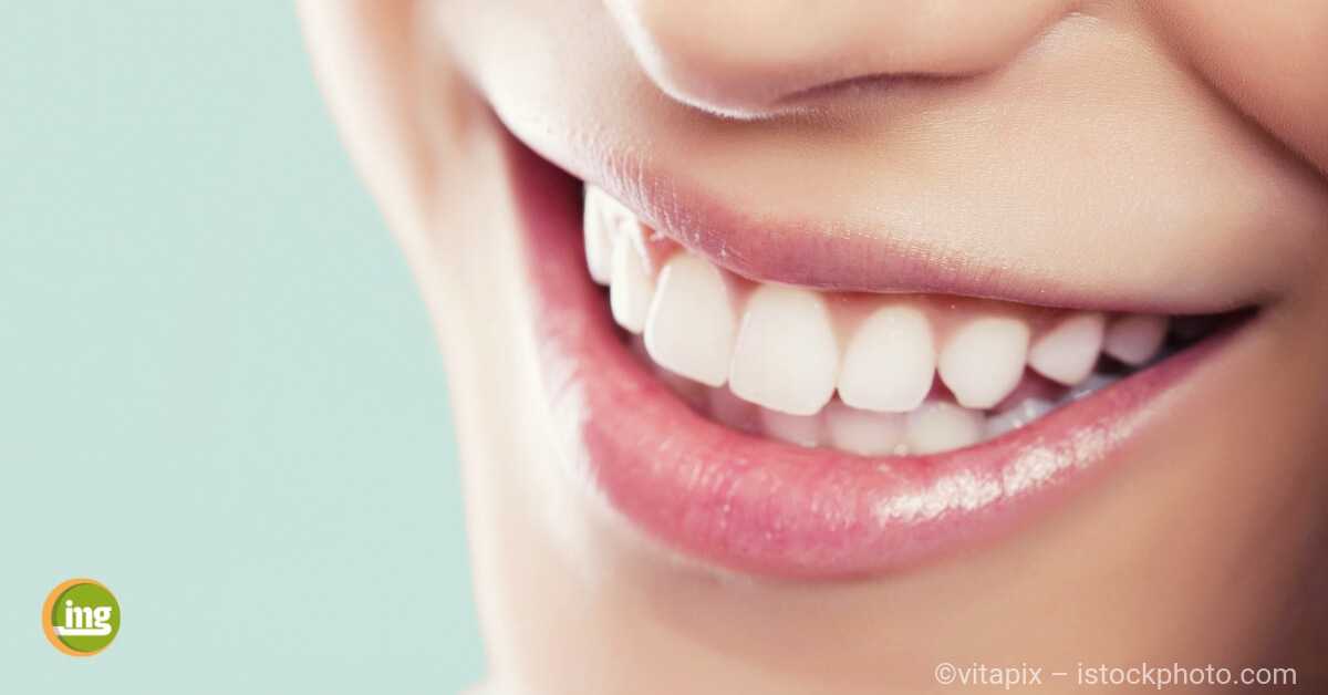
Initial Findings: Oral Carcinoma Photo: K.-D. Wolff-TU Munich
Klaus-Dietrich Wolff et al.
About 10,000 new cases / year develop in the oral cavity, which is five percent of all malignant tumors. With a share of 95 percent, these are predominantly squamous cell carcinomas, which are often associated with the risk factors of chronic alcohol abuse and tobacco consumption [Talamini et al., 2003]. Also, the detection of human papilloma virus (HPV 16) in serum may be a risk factor for oral and pharyngeal carcinoma [Herrero et al., 2003]. Especially men (around 7,500 / year), but increasingly also women are affected; In males, this disease is in the seventh place of all malignant tumors. Although an increase in incidence is registered worldwide, the public perception of this problem is low [Cruz et al., 2002]. Furthermore, there are often differing views on the type and extent of diagnosis, therapy and follow-up. This resulted in the unmistakable need for an S3 guideline, in which the scientific evidence and the practical care of patients with oral cancer in Germany were traceably analyzed, weighed, evaluated and given clear recommendations.
methodology
The guideline was prepared with methodological support from the DKG Guideline Program Oncology. In a kick-off meeting, the guideline group first defined 37 issues relevant to diagnosis, therapy and after-care, which were then systematically analyzed in five working groups and with the professional support of the “Division of Evidence-based Medicine” of the Charité Berlin Literature research with evidence processing were answered. Furthermore, research was carried out on already published guidelines; Due to their timeliness and methodological structure, the SIGN (Scottish Intercollegiate Giudelines Network) 90 guideline was selected as the source guideline. From around 3 000 relevant abstracts, around 250 papers were identified, of which 117 were relevant for further analysis. The literature on which the adapted SIGN-90 guideline was based was compared with the results of the independent de novo research and reassessed in terms of its evidence. Each study was assessed with an evidence level of 1 ++ (high quality meta-analyzes) to 4 (expert opinion). In a concluding consensus conference, all key questions initially defined were answered on the basis of the research carried out and agreed in the form of a nominal group process.
Results
The following statements have been formulated with three levels of recommendation, where “should” is a strong recommendation, “should” be a recommendation and “may” an open recommendation. They summarize the LL’s recommendations without reproducing them verbatim.
All patients with more than two weeks of unclear mucosal lesions should be referred immediately to a specialist for clarification. Suspected is any alteration of the oral mucosa with tissue excess and / or tissue defect as well as a color change or hardening of the mucosa. Typical is a central ulcer with peripheral rim and whitish (leukoplakic) deposits due to cornification (keratinization), which may be completely absent. Early findings are presented, for example, as a nodular epithelial thickening or a flat surface defect. Already initially there may be a tooth loosening or a lymph node swelling on the neck, which can be confused with inflammatory diseases, such as a periodontal disease or a lymphadenitis. As a basic dental diagnosis, a panoramic tomogram should also be available for assessing dental status, also with a view to possible radiotherapy. There is no established evidence for the added benefit of Cone Beam CT (DVT) over panoramic tomography for assessment of mandibular bone invasion [Hendrikx et al., 2010]..
For the initiation of a tumor-specific therapy, the tumor detection by obtaining a histology is a prerequisite. Since the biopsy results in a local tissue reaction that may distort contrast agent imaging (CT, MRI), sampling with clinically apparent tumor findings is recommended only after imaging with contrast agent imaging. The removal of the tissue sample should be done by the specialist from the progression zone of the tumor, ie from its edge area, not from the necrotic center. The usual form of biopsy extraction is incision biopsy with a scalpel. In the case of a brush biopsy care must be taken that it is sufficiently deep with removal of coherent tissue particles and provocation of bleeding to avoid false negatives.
The treatment of oral cancer is to be carried out interdisciplinarily after coordination of each individual case within tumor boards involving the disciplines of oral and maxillofacial surgery, otorhinolaryngology, radiotherapy, oncology, pathology and radiology. To determine the dental status, the examination should be carried out by an experienced dentist before the start of treatment. The therapy should consider the individual situation of the patient. If the general condition of the patient permits it, the operation should be performed on curatively resectable oral carcinoma. Here, reconstructive measures should in principle be part of the surgical concept. The reconstruction should be planned taking into account the overall oncological situation. The expense of the reconstruction should be justified by the expected functional or aesthetic improvement. A decision on surgical therapy should be made taking into account the availability of tumor-free resection margins and the postoperative quality of life. It should be noted that a distance of less than one millimeter between the histologically detectable tumor border and the resection margin is considered a positive cut margin [McMahon et al., 2003]. A resection with a histologically confirmed safety margin of one to three millimeters is referred to as a scarce resection margin, a resection with at least five millimeters as a safe resection margin. Tumor-related or scarce resection margins significantly worsen the prognosis [Loree et al., 1990] Although it has not been possible to prove an improvement in the prognosis by using rapid-cut histology for intraoperative assessment of the tumor borders, this method is uncontrolled or unavoidably more radical in avoiding it Resections helpful [Ribeiro et al., 2003]. It can therefore be assumed that the intraoperative frozen section histology makes a significant contribution to the protection of an R0 resection and to the preservation of structure and function. The continuity of the lower jaw should be preserved during tumor resection, as long as neither imaging nor intraoperative evidence of tumor invasion into the bone could be provided.
An integral part of the treatment of oral carcinoma is the treatment of cervical lymph nodes, which – depending on the preoperative diagnostics (ultrasound, CT, MRI) – can be classified as clinically unobtrusive, suspicious or highly suspicious for tumor invasion. However, it must be taken into account in the treatment decision that histologically, 20 to 40 percent of occult metastases are also found clinically and in the imaging of inconspicuous findings [Coatesworth et al., 2003; Byers et al., 1988]. Therefore, even in patients with clinically unremarkable lymph node status, a selective neck dissection should be performed regardless of the T-category.
While there is no indication for chemoradiotherapy in initial stages or small oral carcinomas, advanced operable carcinomas (T3 / T4) of the oral cavity should be treated with a combination of surgery and chemoradiotherapy. Postoperative radio- or chemoradiotherapy should continue to be performed with scarce or positive resection margins, perineural invasion, vascular invasion and / or lymph node involvement. Postoperative radiotherapy should be started as early as possible and stopped within a maximum of 11 weeks after the operation [Ang et al., 2001]. The indication for primary radiotherapy is given when complete tumor resection can not be achieved or when the surgery would result in significant impairment of quality of life. In patients with advanced, non-operable and non-metastatic oral carcinoma, primary radiochemotherapy should be preferred to radiotherapy alone, especially in those aged up to 70 years [Pignon et al., 2009] To reduce the toxicity of radiotherapy, but none at the same time With the onset of deterioration in local tumor control or overall survival, Intensity Modulated Radiation Therapy (IMRT) has been introduced for patients with head and neck cancers. The aim of avoiding radiation-induced xerostomia by reducing the dose to the salivary glands was achieved in the first series of cases [Chao et al., 2001]. If residual salivary glands remain, oral administration of pilocarpine (5 to 10 mg three times daily) improves oral dryness and reduces the need for artificial saliva [Fisher et al., 2003]..
In the treatment of oral cancer, early dental care is essential to counteract the otherwise commonly occurring tooth loss or radiation caries in the case of planned or previous radiotherapy; This also includes the preparation of a fluoridation and, if appropriate, a spacer bar before the beginning of radiation therapy. Since complications following tooth extraction can be a significant problem, it is imperative that any intervention on the irradiated jaw be performed exclusively by appropriately trained peers with surgical expertise [Epstein et al., 1998]. The most serious complication is infected osteoradione necrosis. Its average incidence is reported as five percent [Tong et al., 1999]. The risk of osteo-radionecrosis increases even further when irradiation for tumor invasion into the jawbone has occurred. Most commonly affected is the molar region of the mandible, and often the infected osteoradion necrosis has been preceded by tooth extraction. Radiation therapy of tumor recurrence with total doses greater than 60 Gy is often responsible for infected osteoradial necrosis in 20% of cases, often in conjunction with chemotherapy [Pasquier et al., 2004]. The treatment of this complication ranges from a systemic antibiotic therapy to the removal of the infected bone and a sequestrotomy to the continuity resection of the affected jaw section, whereby the subsequent reconstruction due to previous operations and radiation is technically demanding. There is insufficient evidence for the benefit of hyperbaric oxygen therapy to prevent or treat osteoradione necrosis. A multicenter case-control study did not show any benefit of hyperbaric oxygen therapy in patients with osteoradionic necrosis when performed without further surgical interventions [Annane et al., 2004].
Treatment of tumor recurrence:
The most frequent reason for an unsuccessful primary tumor treatment and subsequently a tumor-related dying is the locoregional tumor recurrence; It occurs in oral carcinoma in about one fifth of patients. Curative therapeutic options in these cases include reoperation (salvage surgery) [Goodwin, 2000] or / and radio- or chemoradiotherapy [Haraf et al., 1996]. The decision on the appropriate course of action should be based on the patient’s individual situation, the stage of tumor recurrence and its potential resectability, the previous treatment, the likely effectiveness of the therapy, considering its risks and quality of life, general physical condition and not least the patient’s desire must be taken into account. Therapy decision should be made by the interdisciplinary team of the tumor board after histological confirmation of recurrence and re-staging. The patients and their relatives should be informed in detail about the risks of treatment and the prospects of success of the new surgical or conservative therapy, also with regard to a permanent cure, especially with regard to the expected quality of life. The procedure should only be performed by an experienced surgical team with extensive reconstruction options and in a facility with a suitable intensive care facility. Secondary irradiation should ideally be done within a clinical trial trial.
In patients with advanced, relapsed, or metastatic head and neck cancer without curative treatment, response rates of 10 to 35 percent can be achieved with palliative chemotherapy [Schornagel et al., 1995]. Although the response rate of palliative chemotherapy can be improved by a combination of different cytotoxic agents, there is no evidence for prolonged survival [Gibson et al., 2005]. The improvement of the response rate by combination chemotherapy is accompanied by increased hematological and also general toxicity. As with chemotherapy, there are no evidence-based studies available for palliative radiotherapy that can support the efficacy of this treatment modality for incurable head and neck cancer. Patients with incurable tumor disease have a variety of physical and psychological complications that present an additional challenge to treatment. For this reason, these patients should be given early to a professionally carried out supportive therapy.
It is well documented that approximately 90 percent of patients with carcinoma of the oral cavity also suffer from caries, periodontal disease or oral mucosa infection [Rosenberg, 1990], but are often unaware of the need for dental treatment [Toljanic et al. , 2002]. Even under the optimal conditions of continuous dental care, the patient’s ability to be guided can be problematic, as 51 percent of them discontinue aftercare over time [Epstein et al., 1998]..
Since patients are functionally handicapped after tumor-related tooth and / or jaw removal and have a significantly lower quality of life than prosthetically treated patients [Allison et al., 1999], the organization of dental rehabilitation is an important task of tumor aftercare. The prosthetic restoration can be problematic due to the postoperatively altered anatomy and often requires a special commitment of the practitioner. Although the insertion of dental implants in the remaining jawbone or in microvascular anastomosed bone grafts has led to a considerable expansion of the prosthetic possibilities, an increased implant loss rate in the irradiated bone – especially in smokers – must be expected [Mericske-Stern et al. , 1999]. There is insufficient evidence for the most suitable prosthetic procedure in patients who have been operated and / or irradiated for oral cancer [McCord et al., 2004]. Regarding implantation after radiotherapy of the head and neck region, reference is made to the S3 guideline “Implant restoration for oral rehabilitation in connection with head and neck radiation” (AWMF 007-089)
Conclusion for the dental practice
The dental practice is of crucial importance for the detection of oral carcinoma, therapy support and functional rehabilitation. Especially with irradiated patients, a high commitment to avoid complications to hard tooth substance, periodontal and jawbone is required. Dentists have a key role to play in improving the success of oral cancer through early detection.
Summary
In order to make progress in the treatment of oral cancer, a clinical guideline was developed for the first time in collaboration with 21 professional societies and consortiums and with the support of the German Cancer Society at the highest level of evidence (S3). Based on de novo research, systematic reviews, meta-analyzes and taking into account an evidence-based source guideline (SIGN 90), 71 statements and recommendations for the diagnosis and treatment of oral carcinoma were formulated. Explanations in the background text of the guideline also make it possible for third-party colleagues to inform patients about the procedures of the specialists and to advise them on side effects or risks of the therapy. Consistent use is expected to improve the prognosis of affected patients.
Related Posts
-

The most important tool of oral hygiene is still the toothbrush. Unfortunately, it is far too rarely changed by most people. At the latest when bends…
-
Bleaching: therefore only with the dentist, teeth whitening, information oral health
Bleaching – safe and successful only in the dental office! Too much coffee, tea, red wine and nicotine – and the naturally bright color of the teeth…
-

Fixed dentures: for whom are dental implants suitable, information oral health
Safe and solid dentures: For whom are dental implants suitable?? The replacement for the missing tooth in the jaw is called: dental implant. The…
-

No fear of the dentist, information oral health
Do not be afraid of the dentist! Regular visit to the dental office is more than just checking the teeth and gums. Too much health, fitness and…