Supposedly simple movements also result from a complicated interplay of muscles, nerves and parts of the brain. The numerous motor disorders, among which people le > Scientific supervision: Prof. Dr. Jörn Munzert
- Numerous disruptions can occur when planning and executing movements.
- The most common disorders include paralysis due to spinal cord injuries. The height at which the injury happens decides >
spinal cord
Spinal cord / medulla spinalis / spinal cord
The spinal cord is the part of the central nervous system that lies in the spine. It has both the white substance of the nerve fibers and the gray substance of the cell nucleus. Simple reflexes such as the hamstring reflex are already processed here, since sensory and motor neurons are directly connected. The spinal cord is divided into cervical, thoracic, lumbar and sacral marrow.
cerebellum
The cerebellum (cerebellum) is an important part of the brain, located on the back of the brain stem and below the occipital lobe. It consists of two cerebellar hemispheres that are covered by the cerebellar cortex (cerebellar cortex) and plays an important role in automated motor processes, among other things.
inhibition
The neuronal inhibition, or inhibition, describes the phenomenon that a transmitter neuron sends a pulse to the receiver neuron, which leads to a reduction in its activity. The most important inhibitory messenger substance is GABA.
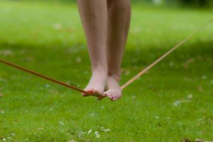
Dancing, swimming, jumping over hurdles or doing a pirouette on the ice are part of the abilities of the human body that require respect from many people. But even for supposedly very simple movements, you need tremendous coordination – even if most people are not aware of it, for example when they walk or lift a drinking glass.
A movement is initially planned in the frontal lobe, more precisely: in the premotor cortex and the so-called supplementary motor area. The actual execution is then initiated in another area of the cerebral cortex, the motor cortex. From there, the commands are sent via nerve fibers in the spinal cord to the muscles that perform the movements. The muscles are constantly monitored by the cerebellum, which compares planned and actual movement – and under certain circumstances intervenes to correct it. The basal ganglia are also involved in motion control. If you watch small children, they learn to stand on their own feet and grope around with the first awkward steps, suspect how great this achievement is. Show people with motor disorders, how much with this complex interplay of different Areas of the brain, nerves and muscles can go wrong.
frontal lobe
Frontal lobe / frontal lobe / frontal lobe
The frontal cortex is the largest of the four lobes of the cerebral cortex and its functions are correspondingly extensive. The front area, the so-called prefrontal cortex, is responsible for complex action planning (so-called executive functions), which also shapes our personality. Its development (myelination) takes up to 30 years and is still not yet complete. Other important components of the frontal cortex are the Broca area, which controls our linguistic expression, and the primary motor cortex, which sends movement impulses to the entire body.
Cortex
Cerebral cortex / cortex cerebri / cerebral cortex
The cortex cerebri, or cortex for short, denotes the outermost layer of the cerebrum. It is 2.5 mm to 5 mm thick and rich in nerve cells. The cerebral cortex is strongly folded, comparable to a handkerchief in a cup. This creates numerous turns (gyri), crevices (fissurae) and furrows (sulci). When unfolded, the surface of the cortex is approximately 1,800 cm 2 .
spinal cord
Spinal cord / medulla spinalis / spinal cord
The spinal cord is the part of the central nervous system that lies in the spine. It has both the white substance of the nerve fibers and the gray substance of the cell nucleus. Simple reflexes such as the hamstring reflex are already processed here, since sensory and motor neurons are directly connected. The spinal cord is divided into cervical, thoracic, lumbar and sacral marrow.
cerebellum
The cerebellum (cerebellum) is an important part of the brain, located on the back of the brain stem and below the occipital lobe. It consists of two cerebellar hemispheres that are covered by the cerebellar cortex (cerebellar cortex) and plays an important role in automated motor processes, among other things.
basal ganglia
Basal ganglia / nuclei basales / basal ganglia
Basal ganglia are a group of subcortical nuclei (located below the cerebral cortex) in the telencephalon. The basal ganglia include the globus pallidus and the striatum. Some authors include other structures, such as. B. the Claustrum. The basal ganglia are primarily associated with voluntary motor skills.
Paralysis can have many causes
The most common motor disorders include paralysis, in which those affected can no longer move individual muscles, parts of the body or the whole body. Many paralyzes are caused by spinal cord injuries. One of the most severe and well-known forms is paraplegia, which occurs in around 1,000 people in Germany every year. The spine is damaged so badly – usually by an accident – that the nerve fibers that run through it are cut or squeezed and the signals from the brain can no longer be passed on to the muscles.
Depending on the level at which this interruption occurs, patients are paralyzed to different degrees. "The higher the spine the injury is, more more muscles are affected by the paralysis, ”says Gabriel Curio, neurologist at the Charité, the Berlin University Hospital. A cross-section below C4 means that the fourth of the seven vertebral bodies of the cervical spine is still intact. Such a patient can still control the diaphragm and thus his breathing. If the injury is further up, for example at the level of the second cervical vertebra, the patient must be artificially ventilated. Patients with a thoracic vertebra can also move their arms and hands, but have difficulty moving their upper body because they have no control over their abdominal muscles.
In addition to accidents, herniated discs can also lead to such paralysis. Intervertebral discs serve as a buffer between the vertebral bodies, explains Curio. "If they are squeezed too much, it may be that the connective tissue tape that runs around the disc tears and the gelatinous mass slides aside." Then the nerve gets trapped and cannot transmit the stimuli from the brain either.
spinal cord
Spinal cord / medulla spinalis / spinal cord
The spinal cord is the part of the central nervous system that lies in the spine. It has both the white substance of the nerve fibers and the gray substance of the cell nucleus. Simple reflexes such as the hamstring reflex are already processed here, since sensory and motor neurons are directly connected. The spinal cord is divided into cervical, thoracic, lumbar and sacral marrow.
RELATED ITEMS
-
Exercise, sport and fun – but safe – sports for children from a-z
Martina Abel and Inke Ruhe It is becoming increasingly important to get children moving, to actively offer them opportunities to move. This also includes the…
-
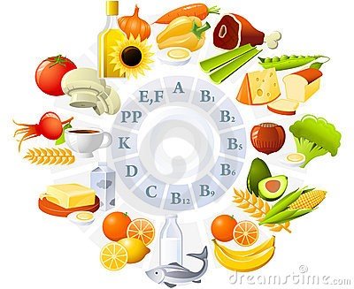
The combination of exercise and nutrition … .. Nutrition and exercise are two of the most important building blocks for a healthy life. A child who cares a lot…
-

Children and movement – why movement is so important, Germany
Movement invigorates and enables a healthy and vital life. It is not always the top athletic performance that our body needs…
-

At what age do babies learn to sit
Nicole Wendler holds a doctorate in biology from the field of oncology and immunology. As a medical editor, author and editor, she is for different people…