
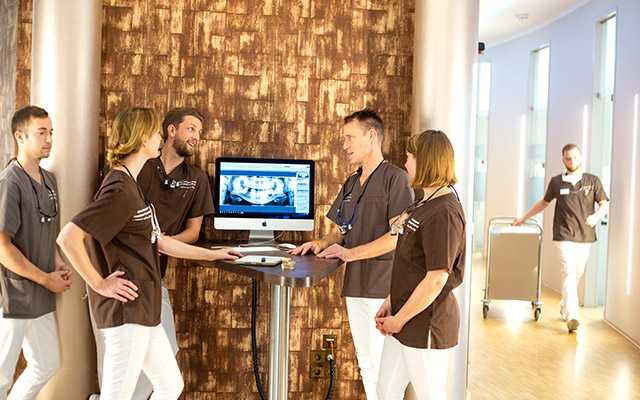
High tech in dentistry
In our clinic, the treatments are carried out with the most modern dental technology, the diagnostic work, the practical experience and the expertise of our doctors are effectively supported by the technical infrastructure.
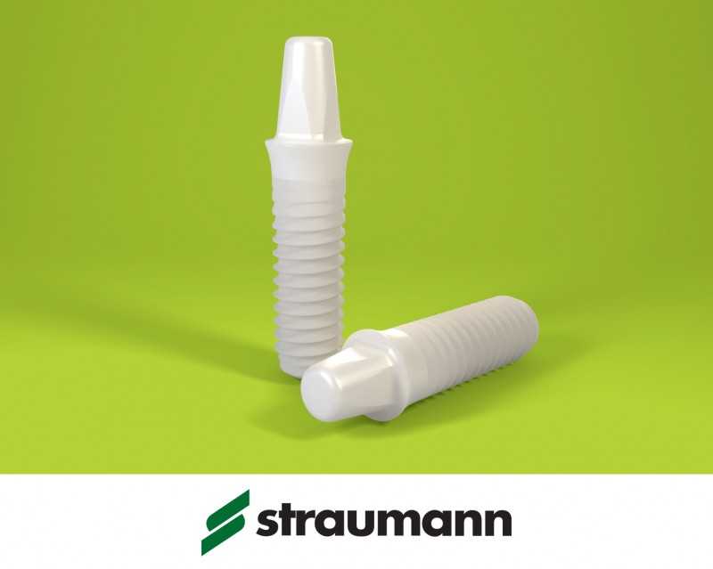
The Gelencsér Dental Dental Clinic does its best to ensure the safety and satisfaction of its patients: computer-assisted 3D tomography, digital x-ray diagnostics, state-of-the-art implant technology, laser surgery and the latest dental bleaching procedures provide patients with accurate and successful therapies.
The whole thing is rounded off by the know-how and routine of our dentists, who in recent years have expanded their expertise by treating foreign patients.
Our specialists have excellent training at renowned Hungarian medical universities and are still striving to keep their knowledge up to date and to acquire innovative techniques.
For their professional development, they regularly take part in various representative medical conferences in Germany and abroad.
3D planning, effective solutions
The dental clinic has 21st-century, 3-dimensional medical devices that enable accurate and perfect diagnoses, effective dental treatments, and a speedy recovery.
Using state-of-the-art technologies such as 3D computed tomography, 2-dimensional panoramic x-ray and intraoral camera, our specialists are able to get an accurate picture of the condition of the patient’s teeth at check-up.
Our 10 treatment rooms are air-conditioned and equipped with modern treatment chairs.
Thanks to the targeted use of appropriate anaesthesiological techniques, the treatments are completely painless, but at the request of the patient they can also be performed in drowsy sleep anesthesia völlig to completely shield acoustic and visual stimuli. We have qualified anaesthesiologists at our disposal.
High-tech in the treatment rooms at the Gelencsér Dental Dental Clinic:
- 3D Cone Beam CT – digital volume tomography
- safe, digital panoramic x-ray with low radiation exposure
- Intraoral camera for even more accurate, detailed diagnostics
- Magnifying glasses and dental microscope for a perfect result
- Functional diagnostics for the optimal fit of dentures
- digital image documentation for easier communication
- 3D CAD / CAM technology for the production of perfect dentures
3D volume tomography
Cutting edge technology and digital imaging at Gelencsér Dental Clinic: Cone Beam CT 3 dimensional digital volume tomography.
Digital Volume Tomography (DVT) is a 3-dimensional imaging technique, i. an imaging tomography procedure. This procedure gives accurate image-diagnostic help using X-rays (with the use of extremely low 68 μSv radiation dose), which is mainly used in dentistry in otorhinolaryngology, oral and maxillofacial surgery, implantology , Periodontology and in orthodontics is used.
The most important applications of Cone-Beam-CT:
- implant design
- Implantation of teeth and implants
- Tooth Herd – Herdtests
- Wisdom tooth analysis
- Examination of the lower jawbone surface
Digital X-ray
Digital X-ray is currently the best and most gentle X-ray examination method. Instead of the previously used X-ray film, Digital X-ray uses a sensor that is extremely sensitive to X-rays.
Thus, the radiation exposure can be reduced by more than 50%, false exposures – and thus multiple X-rays are avoided. We think that’s a great effect for our patients.
The pin-sharp digital images appear on the monitor within a few seconds and can be optimally diagnosed, as they can be enlarged as needed.
Dental laser for more effective solutions
Laser is considered one of the most popular technologies in dentistry, and is used in many areas of modern dentistry best use.
This has the plausible reason that bundled laser beams can also be used in places that were previously inaccessible with the usual dental instruments.
Laser treatments are associated with less pain and are considered an effective, gentle method.
The laser is an indispensable part of innovative dentistry; it not only offers potential for root and gum disease, it also makes it easy to recognize and treat tooth decay.
Laser treatments are a gentle alternative that is also favored by our specialists in various dental preservation procedures as well as aesthetic and oral surgery treatments. During treatment, the patient and the treatment team wear special protective goggles.
Laser technology offers numerous advantages over conventional treatment: laser treatments are characterized by faster wound healing, less stress, a feeling of comfort and minimal bleeding. The result can already be seen after the first treatment.
In which areas is the dental laser used??
- caries treatment
- root canal treatment
- for oral surgery, for cutting of soft tissue with low blood pressure and for the promotion of bone regeneration
- Gum shaping
- in periodontitis ↗
- Curettage / deep cleansing
- tooth whitening
Root canal treatment with dental laser?
Yes! In a root canal treatment, the laser is used primarily for disinfecting the root canal. With the laser, inflamed areas of the dental pulp can be eliminated but the key advantage is that pathogens can be killed even in hard to reach root canals.
How can the dental laser be used in periodontitis treatment?
With the laser light, the bacteria causing bacteria in the root canals can be killed without supplying the body with harmful drugs. With this completely painless procedure even the deep under the gum nesting germs, which are responsible for the inflammation, to eliminate.
Dental laser against tooth decay?
Thanks to the laser caries can be removed without drilling, without pain and without anesthetic injection. This procedure is much more effective than the traditional method of treatment, but more importantly, it is more comfortable and completely stress-free for the patient.
Are also sensitive teeth to be treated with laser?
The sensitivity of the tooth necks can be alleviated by lasers again.
In which areas can the dental laser still be used sensibly?
The dental laser is characterized by its wide range of applications and is in both oral surgery, as well as aesthetic and conservative dental treatments of great benefit.
Intraoral camera
Digital image processing dominates our everyday lives, and does not stop at dental treatment rooms. Multimedia technology is meeting more and more dentists who prefer to use an exceptional device to support patient communication.
The intraoral camera provides transparency and provides a revolutionary, objective method for optimal patient education. When patients see their teeth from the perspective of the dentist, they can directly understand the need for treatment.
This intra-oral camera is basically a small camera whose images are displayed on a screen during treatment so that the patient can see with his own eyes what the dentist is talking about.
Intraoral cameras are becoming ever more flexible, small, ultralight, and extremely easy to use. Thanks to LED lighting, they are extremely reliable and versatile, even in hard-to-reach places in the mouth. The images can be archived for later work phases with a simple click.
Diagnoses have much more informative value in image format and create a good basis of trust. Everyone understands images, so they also serve in the prophylaxis of an active enlightenment and motivation.
Complementing panoramic X-rays and CT images, these live on-screen images provide better understanding. Nothing provides insight more quickly than when the patient sees what only his dentist sees.
Dental Microscope: more precision, more effective results
A good judgment is required throughout the diagnostics and dental treatment, otherwise it would be very doubtful to recognize caries, dental disease and other diseases in the oral cavity. However, eye-goodness alone is not always enough; one often resorts to precision tools.
For the greatest possible safety and for more effective results, specialists often support their trained eyes with high-tech achievements, according to the motto "More quality through more vision".
Magnifying glasses for work control
The loupes provide an excellent opportunity to check the procedures and laboratory models in order to guarantee precision and durability when checking the performed dental work.
Dental microscope for the detail accuracy
The high-magnification dental microscope is primarily used in the dental laboratory, where colleagues can review the images on a high-resolution monitor and rely on this detailed information as they work. This instrument provides solid help to ensure the best results and quality.
Caries detector for more precise diagnoses
The caries detector is actually a chemical indicator test that can also reveal hidden caries in order to be able to cope with this problem in good time.
Functional diagnostics: exact, precise analysis, perfect results
Although physical examination plays an important role in diagnostics, in the majority of cases physicians rely on instrumental functional diagnostics, which makes it easy to identify malfunctions in the dentition system.
Functional diagnostics provide accurate information about whether each treatment can be performed and help to create a perfectly functioning, long-lasting denture.
Long-lasting crowns, bridges, prostheses and bite splints can be precisely planned and manufactured thanks to this diagnostic procedure, since functional prostheses can only be adapted after having been informed of precise information.
This helps to prevent future diseases of the temporomandibular joints and the patient leaves our clinic with a perfectly fitting prosthesis.
How to determine the necessary data in the instrumental functional diagnostics?
The measurement is completely painless and takes place under relaxed circumstances. The specialist uses the measuring device, the so-called facebow, at the corresponding measuring points in order to obtain an impression of the upper and lower jaw.
The facebow measures the individual positional relationship of the upper jaw to the skull and joints, so the dental technicians from the impression can make the identical model of your jaw.
The measurement data are recorded in a special articulator, which is able to mimic the natural movements of the modeled jaw.
The simulation provides dentists and dental technicians with essential information on the preparation of dentures and bite splints with a perfect fit.
Digital photo documentation
We document your entire course of treatment through digital photography – at the beginning, during and after the completion of your treatment.
The photo documentation ensures a high degree of communication between dentist and laboratory, but also for you as a patient, the pictorial illustration is interesting:
Based on the pictures, we can illustrate the success of the treatment by comparing the initial and final situation. Many of our patients are even more motivated to sustain the positive result over the long term with our support.
Thanks to the digital networking of our dental practice, the dental technology team can also access the image files if necessary.
Related Posts
-
Dental practice – dental clinic hannover, surgery dentistry
Dental practice and dental clinic in Hanover: surgery for teeth and gums The PODBI344 is also well equipped for the more complex cases! For surgical…
-
Dental Implants: Costs, Manufacturers, Experiences and Treatment
Everything about dental implants There are certainly many questions that you would like clarified if you lose one or more teeth and this through Dental…
-
Dentistry herne »holistic dental treatment
dentistry Holistic Dentistry in the Dental Clinic Herne The term dentistry is often used as a comprehensive synonym for oral and maxillofacial dentistry….
-
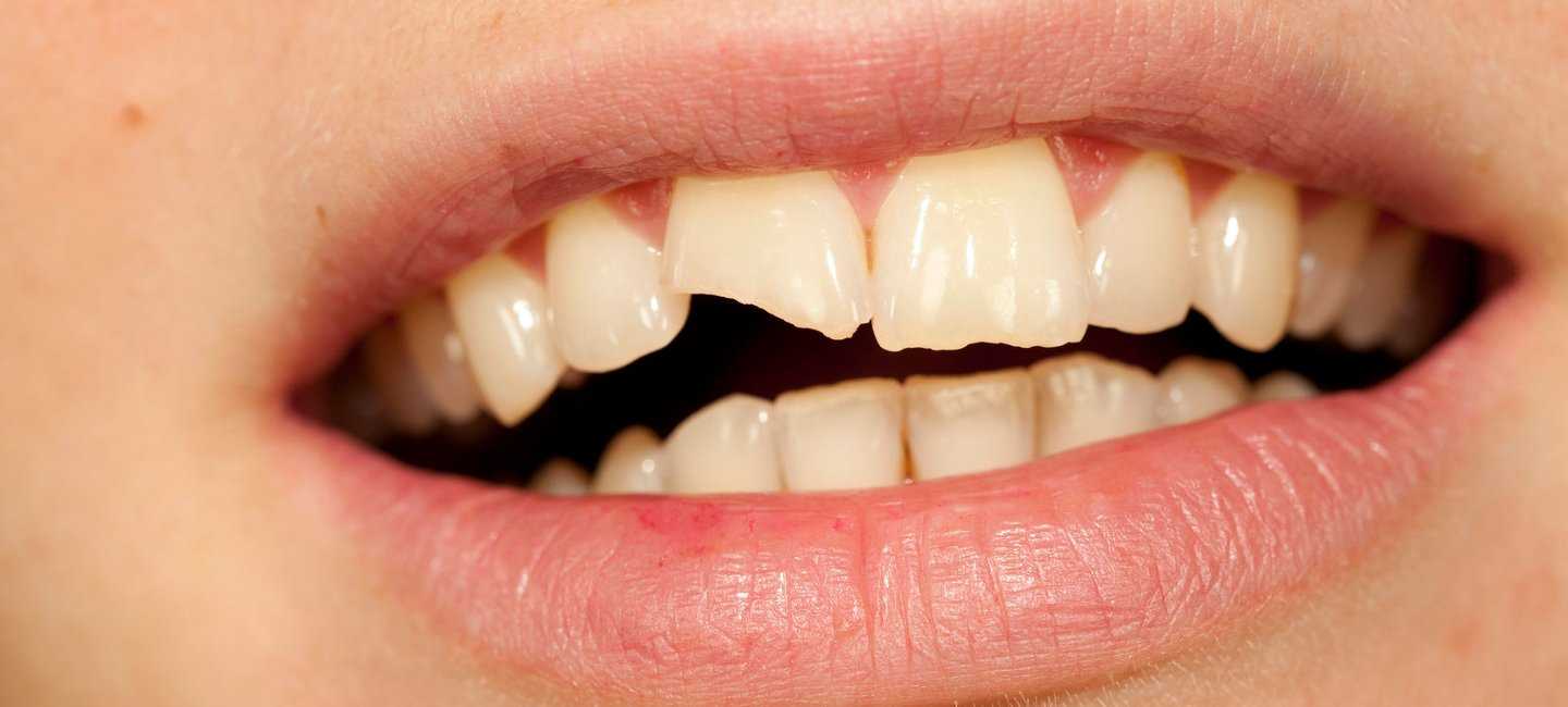
Dental accident – what to do, center for dentistry
Dental accident – What to do if the tooth has broken off? A dental accident is a very painful and unpleasant experience. Children and adolescents are…